Simulating shared segments between relatives

A few months ago I saw this nice figure from Amy Williams of the number of DNA segments that are expected to be shared between relatives. I thought it would be fun to simulate segment sharing with AlphaSimR.

Because DNA comes in chromosomes that don’t break up and recombine that much, the shared DNA between relatives tends to come in long chunks — segments that are identical by descent. The distribution of segment lengths can sometimes be used to tell apart relationships that would otherwise give the same average (e.g., Yengo et al. 2019, Qiao et al. 2021).

But let’s not do anything sophisticated. Instead, we take three very simple pedigrees — anyone who’s taken introductory genetics will recognize these ones — and look at relationships between full-sibs, half-sibs and cousins. We’ll also look at the inbred offspring of matings between full-sibs, half-sibs and cousins to see that the proportion that they share between their two copies of the genome lines up with the expected inbreeding.

There won’t be any direct comparison to the values that Williams’ simulation, because it simulated more distant relationships than this, starting with cousins and then moving further away. This is probably more interesting, especially for human genealogical genetics.

The code is on GitHub if you wants to follow along.

The pedigrees

Here are the three pedigrees, drawn with the kinship2 package:

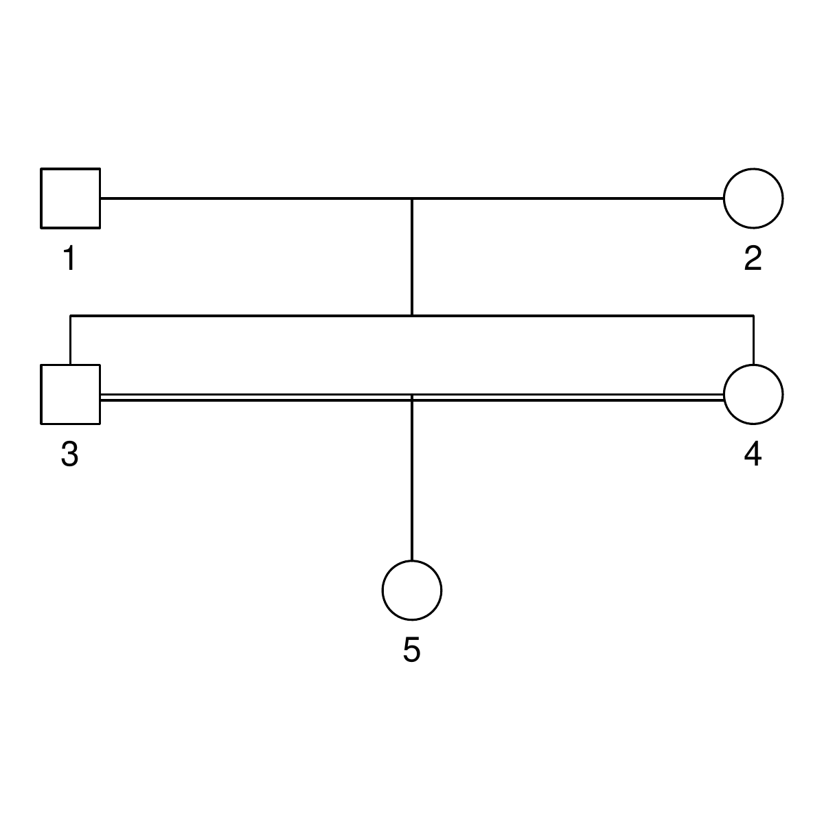
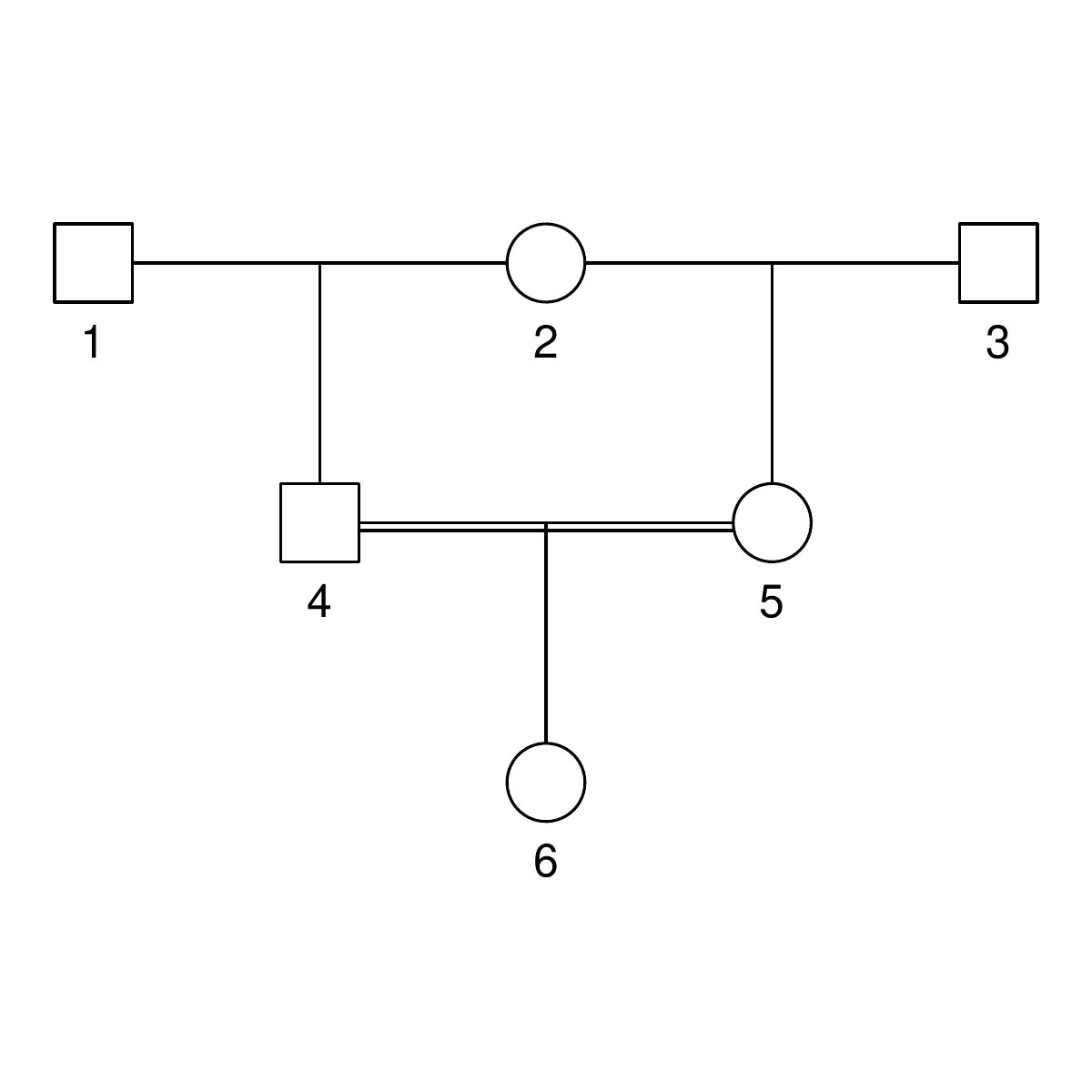
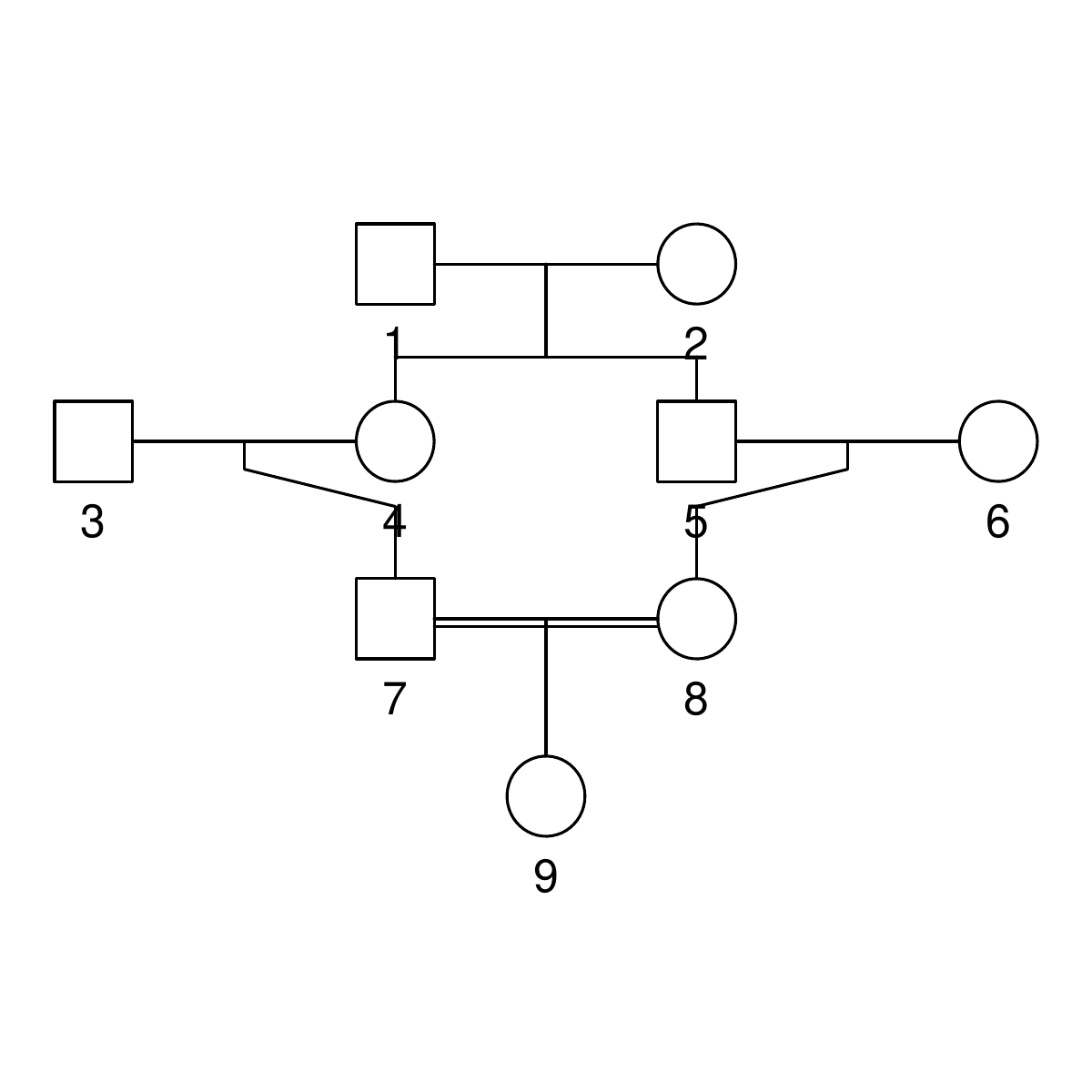
A pedigree, here, is really a table of individuals, where each column tells us their identifier, their parents, and optionally their sex, like this:

id, mother, father, sex
1, NA, NA, M
2, NA, NA, F
3, NA, NA, M
4, 2, 1, F
5, 2, 1, M
6, NA, NA, F
7, 4, 3, M
8, 6, 5, F
9, 8, 7, F

We can use GeneticsPed to check the relatedness and inbreeding if we don’t trust that I’ve entered the pedigrees right.

library(GeneticsPed)
library(purrr)
library(readr)

ped_fullsib <- read_csv("pedigrees/inbreeding_fullsib.txt")
ped_halfsib <- read_csv("pedigrees/inbreeding_halfsib.txt")
ped_cousin <- read_csv("pedigrees/inbreeding_cousin.txt")

inbreeding_ped <- function(ped) {

    inbreeding(Pedigree(ped))

}

print(map(list(ped_fullsib, ped_halfsib, ped_cousin), inbreeding_ped))
[[1]]
   1    2    3    4    5 
0.00 0.00 0.00 0.00 0.25 

[[2]]
    1     2     3     4     5     6 
0.000 0.000 0.000 0.000 0.000 0.125 

[[3]]
     1      2      3      4      5      6      7      8      9 
0.0000 0.0000 0.0000 0.0000 0.0000 0.0000 0.0000 0.0000 0.0625

Comparing haplotypes

We need some functions to compare haplotypes and individuals:

library(AlphaSimR)
library(dplyr)
library(purrr)
library(tibble)

## Find shared segments between two haplotypes expressed as vectors
## map is a vector of marker positions

compare_haplotypes <- function(h1, h2, map) {
    sharing <- h1 == h2

    runs <- rle(sharing)
    end <- cumsum(runs$lengths)
    start <- c(1, end[-length(end)] + 1)

    segments <- tibble(start = start,
                       end = end,
                       start_pos = map[start],
                       end_pos = map[end],
                       segment_length = end_pos - start_pos,
                       value = runs$values)

    segments[segments$value,]
}

We will have haplotypes of the variants that go together on a chromosome, and we want to find segments that are shared between them. We do this with a logical vector that tests each variant for equality, and then use the rle to turn this into run-length encoding. We extract the start and end position of the runs and then keep only the runs of equality.

Building on that function, we want to find the shared segments on a chromosome between two individuals. That is, we make all the pairwise comparisons between the haplotypes they carry and combine them.

## Find shared segments between two individuals (expressed as
## matrices of haplotypes) for one chromosome

compare_individuals_chr <- function(ind1, ind2, map) {

    h1_1 <- as.vector(ind1[1,])
    h1_2 <- as.vector(ind1[2,])

    h2_1 <- as.vector(ind2[1,])
    h2_2 <- as.vector(ind2[2,])

    sharing1 <- compare_haplotypes(h1_1, h2_1, map)
    sharing2 <- compare_haplotypes(h1_1, h2_2, map)
    sharing3 <- compare_haplotypes(h1_2, h2_1, map)
    sharing4 <- compare_haplotypes(h1_2, h2_2, map)

    bind_rows(sharing1, sharing2, sharing3, sharing4)
}

Finally, we use that function to compare individuals along all the chromosomes.

This function takes in a population and simulation parameter object from AlphaSimR, and two target individuals to be compared.

We use AlphaSimR‘s pullIbdHaplo function to extract tracked founder haplotypes (see below) and then loop over chromosomes to apply the above comparison functions.

## Find shared segments between two target individuals in a
## population

compare_individuals <- function(pop,
                                target_individuals,
                                simparam) {

    n_chr <- simparam$nChr

    ind1_ix <- paste(target_individuals[1], c("_1", "_2"), sep = "")
    ind2_ix <- paste(target_individuals[2], c("_1", "_2"), sep = "")

    ibd <- pullIbdHaplo(pop,
                        simParam = simparam)

    map <- simparam$genMap
    loci_per_chr <- map_dbl(map, length)

    chr_ends <- cumsum(loci_per_chr)
    chr_starts <- c(1, chr_ends[-n_chr] + 1)

    results <- vector(mode = "list",
                      length = n_chr)

    for (chr_ix in 1:n_chr) {

        ind1 <- ibd[ind1_ix, chr_starts[chr_ix]:chr_ends[chr_ix]]
        ind2 <- ibd[ind2_ix, chr_starts[chr_ix]:chr_ends[chr_ix]]

        results[[chr_ix]] <- compare_individuals_chr(ind1, ind2, map[[chr_ix]])
        results[[chr_ix]]$chr <- chr_ix
    }

    bind_rows(results)
}

(You might think it would be more elegant, when looping over chromosomes, to pull out the identity-by-descent data for each chromosome at a time. This won’t work on version 1.0.4 though, because of a problem with pullIbdHaplo which has been fixed in the development version.)

We use an analogous function to compare the haplotypes carried by one individual. See the details on GitHub if you’re interested.

Building the simulation

We are ready to run our simulation: This code creates a few founder individuals that will initiate the pedigree, and sets up a basic simulation. The key simulation parameter is to set setTrackRec(TRUE) to turn on tracking of recombinations and founder haplotypes.

source("R/simulation_functions.R")

## Set up simulation

founders <- runMacs(nInd = 10,
                    nChr = 25)

simparam <- SimParam$new(founders)

simparam$setTrackRec(TRUE)

founderpop <- newPop(founders,
                     simParam = simparam)

To simulate a pedigree, we use pedigreeCross, a built-in function to simulate a given pedigree, and then apply our comparison functions to the resulting simulated population.

## Run the simulation for a pedigree one replicate

simulate_pedigree <- function(ped,
                              target_individuals,
                              focal_individual,
                              founderpop,
                              simparam) {
    pop <- pedigreeCross(founderPop = founderpop,
                         id = ped$id,
                         mother = ped$mother,
                         father = ped$father,
                         simParam = simparam)
    shared_parents <- compare_individuals(pop,
                                          target_individuals,
                                          simparam)
    shared_inbred <- compare_self(pop,
                                  focal_individual,
                                  simparam)
    list(population = pop,
         shared_segments_parents = shared_parents,
         shared_segments_self_inbred = shared_inbred)
}

Results

First we can check how large proportion of the genome of our inbred individuals is shared between their two haplotypes, averaged over 100 replicates. That is, how much of the genome is homozygous identical by descent — what is their genomic inbreeding? It lines up with the expectation form pedigree: 0.25 for the half-sib pedigree, close to 0.125 for the full-sib pedigree and close to 0.0625 for the cousin pedigree. The proportion shared by the parents is, as it should, about double that.

      case inbred_self_sharing parent_sharing
  full-sib        0.25 (0.052)    0.5 (0.041)
  half-sib        0.13 (0.038)   0.25 (0.029)
    cousin       0.064 (0.027)   0.13 (0.022)

Table of the mean proportion of genome shared between the two genome copies in inbred individuals and between their parents. Standard deviations in parentheses.

This is a nice consistency check, but not really what we wanted. The point of explicitly simulating chromosomes and recombinations is to look at segments, not just total sharing.

With a little counting and summarisation, we can plot the distributions of segment lengths. The horizontal axis is the length of the segments expressed in centimorgan. The vertical axis is the number of shared
segments of this length or longer. Each line is a replicate.

If we look at the summaries (table below), full-sibs share on average 74 segments greater than 1 cM in length, half-sibs 37, and cousins 29.

In real data, short segments might be harder to detect, but because we’re using simulated fake data, we don’t have to worry about phasing errors or false positive sharing.

If we look only at long segments (> 20 cM), full-sibs share on average 46 segments, half-sibs 23, and cousins 13. (Also, similar to Williams’ simulations, none of the cousins simulated here had less than five long segments shared.)

  case     `1 cM`   `10 cM`  `20 cM`  `30 cM`  `40 cM`  
  full-sib 74 (5.2) 60 (4.2) 46 (3.6) 34 (4)   24 (3.8) 
  half-sib 37 (3.4) 30 (3.1) 23 (3.3) 17 (2.8) 13 (2.6) 
  cousin   29 (3.8) 20 (3.3) 13 (3.2) 7.6 (2)  4.3 (1.8)

Table of the mean number of shared segments of different minimum length. Standard deviations in parentheses.

We an also look at the average length of the segments shared, and note that while full-sibs and half-sibs differ in the number of segments, and total segment length shared (above), the length of individual segments is about the same:

  case     mean_length_sd
  full-sib 0.33 (0.032)  
  half-sib 0.34 (0.042)  
  cousin   0.21 (0.03)

Table of the mean length shared segments. Standard deviations in parentheses.

Limitations

Williams’ simulation, using the ped-sim tool, had a more detailed model of recombination in the human genome, with different interference parameters for each chromosome, sex-specific recombination and so on. In that way, it is much more realistic.
We’re not modelling any one genome in particular, but a very generic genome. Each chromosome is 100 cM long for example; one can imagine that a genome with many short chromosomes would give a different distribution. This can be changed, though; the chromosome size is the easiest, if we just pick a species.

Literature

Yengo, L., Wray, N. R., & Visscher, P. M. (2019). Extreme inbreeding in a European ancestry sample from the contemporary UK population. Nature communications, 10(1), 1-11.

Qiao, Y., Sannerud, J. G., Basu-Roy, S., Hayward, C., & Williams, A. L. (2021). Distinguishing pedigree relationships via multi-way identity by descent sharing and sex-specific genetic maps. The American Journal of Human Genetics, 108(1), 68-83.

Estimating recent population history from linkage disequilibrium with GONE and SNeP

In this post, we will look at running two programs that infer population history — understood as changes in linkage disequilibrium over time — from genotype data. The post will chronicle running them on some simulated data; it will be light on theory, and light on methods evaluation.

Linkage disequilibrium, i.e. correlation between alleles at different genetic variants, breaks down over time when alleles are shuffled by recombination. The efficiency of that process depends on the distance between the variants (because variants close to each other on the same chromosome will recombine less often) and the population size (because more individuals means more recombinations). Those relationships mean that the strength of linkage disequilibrium at a particular distance between variants is related to the population size at a particular time. (Roughly, and assuming a lot.) There are several methods that make use of the relationship between effective population size, recombination rate and linkage disequilibrium to estimate population history.

The programs

The two programs we’ll look at are SNeP and GONE. They both first calculate different statistics of pairwise linkage disequilibrium between markers. SNeP groups pairs of markers into groups based on the distance between them, and estimates the effective population size for each group and how many generations ago each group represents. GONE goes further: it uses a formula for the expected linkage disequilibrium from a sequence of effective population sizes and a genetic algorithm to find such a sequence that fits the observed linkage disequilibrium at different distances.

Paper about GONE: Santiago, E., Novo, I., Pardiñas, A. F., Saura, M., Wang, J., & Caballero, A. (2020). Recent demographic history inferred by high-resolution analysis of linkage disequilibrium. Molecular Biology and Evolution, 37(12), 3642-3653.

Paper about SNeP: Barbato, M., Orozco-terWengel, P., Tapio, M., & Bruford, M. W. (2015). SNeP: a tool to estimate trends in recent effective population size trajectories using genome-wide SNP data. Frontiers in genetics, 6, 109.

These methods are suited for estimating recent population history in single closed populations. There are other methods, e.g. the Pairwise Markovian coealescent and methods based on Approximate Bayesian Computation, that try to reach further back in time or deal with connected populations.

(Humorously, barring one capitalisation difference, GONE shares it’s name with an unrelated program related to effective population sizes, GONe … There are not enough linkage disequilibrium puns to go around, I guess.)

Some fake data

First, let us generate some fake data to run the programs on. We will use the Markovian coalescent simulator MaCS inside AlphaSimR. That is, we won’t really make use of any feature of AlphaSimR except that it’s a convenient way to run MaCS.

There is a GitHub repo if you want to follow along.

We simulate a constant population, a population that decreased in size relatively recently, a population that increased in size recently, and a population that decreased in longer ago. The latter should be outside of what these methods can comfortably estimate. Finally, let’s also include a population that has migration from an other (unseen) population. Again, that should be a case these methods struggle with.

Simulated true population histories. Note that the horizontal axis is generations ago, meaning that if you read left to right, it runs backwards in time. This is typical when showing population histories like this, but can be confusing. Also not the different scales on the horizontal axis.

library(AlphaSimR)
library(purrr)
library(tibble)

## Population histories

recent_decrease <- tibble(generations = c(1, 50, 100, 150),
                          Ne = c(1000, 1500, 2000, 3000))

recent_increase <- tibble(generations = c(1, 50, 100, 150),
                          Ne = c(3000, 2000, 1500, 1000))

ancient_decrease <- tibble(generations = recent_decrease$generations + 500,
                           Ne = recent_decrease$Ne)

We can feed these population histories (almost) directly into AlphaSimR’s runMacs2 function. The migration case is a little bit more work because we will to modify the command, but AlphaSimR still helps us. MaCS takes a command line written using the same arguments as the paradigmatic ms program. The runMacs2 function we used above generates the MaCS command line for us; we can ask it to just return the command for us to modify. The split argument tells us that we want two populations that split 100 generations ago.

runMacs2(nInd = 100,
         Ne = recent_decrease$Ne[1],
         histGen = recent_decrease$generations[-1],
         histNe = recent_decrease$Ne[-1],
         split = 100,
         returnCommand = TRUE)

The resulting command looks like this:

"1e+08 -t 1e-04 -r 4e-05 -I 2 100 100  -eN 0.0125 1.5 -eN 0.025 2 -eN 0.0375 3 -ej 0.025001 2 1"

The first part is the number of basepairs on the chromosome, -t flag is for the population mutation rate \theta = 4 N_e \mu, -r for the recombination rate (also multiplied by four times the effective population size). The -eN arguments change the population size, and the -ej argument is for splitting and joining populations.

We can check that these numbers make sense: The population mutation rate of 10-4 is the typical per nucleotide mutation rate of 2.5 × 10⁻⁸ multiplied by four times the final population size of 1000. The scaled recombination rate of 4 × 10⁻⁵ is typical per nucleotide recombination rate of 10-8 multiplied by the same.

The population size changes (-eN arguments) are of the format scaled time followed by a scaling of the final population size. Times are scaled by four times the final population size, again. This means that 0.0125 followed by 1.5 refers to that 4 × 0.0125 × 1000 = 50 generations ago population size was 1.5 times the final population size, namely 1500. And so on.

-I (I assume for ”island” as in the ”island model”) sets up the two populations, each with 100 individuals and a migration rate between them. We modify this by setting it to 200 for the first population (because we want diploid individuals, so we need double the number of chromosomes) and 0 for the other; that is, this ghost population will not be observed, only used to influence the first one by migration. Then we set the third parameter, that AlphaSimR has not used: the migration rate. Again, this is expressed as four times the final population size, so for a migration rate of 0.05 we put 200.

Now we can run all cases to generate samples of 100 individuals.

migration_command <- "1e+08 -t 1e-04 -r 4e-05 -I 2 200 0 200  -eN 0.0125 1.5 -eN 0.025 2 -eN 0.0375 3 -ej 0.025001 2 1"

pops <- list(pop_constant = runMacs2(nInd = 100,
                                     nChr = 5,
                                     histNe = NULL,
                                     histGen = NULL,
                                     Ne = 1000),
             
             pop_recent = runMacs2(nInd = 100,
                                   nChr = 5,
                                   Ne = recent_decrease$Ne[1],
                                   histGen = recent_decrease$generations[-1],
                                   histNe = recent_decrease$Ne[-1]),
             
             pop_increase = runMacs2(nInd = 100,
                                     nChr = 5,
                                     Ne = recent_increase$Ne[1],
                                     histGen = recent_increase$generations[-1],
                                     histNe = recent_increase$Ne[-1]),
             
             pop_ancient = runMacs2(nInd = 100,
                                    nChr = 5,
                                    Ne = ancient_decrease$Ne[1],
                                    histGen = ancient_decrease$generations[-1],
                                    histNe = ancient_decrease$Ne[-1]),
             
             pop_migration = runMacs(nInd = 100,
                                     nChr = 5,
                                     manualCommand = migration_command,
                                     manualGenLen = 1))

Both GONE and SNeP work with text files in Plink ped/map format. Look in the repository if you want to see the not too exciting details of how we extract 10 000 markers and save them to Plink format together with their positions.

Running GONE

GONE source code and binaries are found in their GitHub repository, which we can clone or simply download from the web. As I’m running on Linux, we will use the binaries in the Linux subfolder. Before doing anything else, we will have to set the permissions for each of the binaries stored in the PROGRAMMES directory, with chmod u+x to make them executable.

GONE consists of a set of binaries and a bash script that runs them in order. As the as the script assumes it’s always running from the directory where you put GONE and always writes the output files into the same directory, the easiest way to handle it is to duplicate the entire GONE directory for every run. This also duplicates the INPUT_PARAMETERS_FILE file that one would modify to change settings. Then, invoking GONE with default settings would be as simple as opening a terminal, moving to the working directory and running the script, feeding it the base name of the Plink file:

./script_GONE.sh pop_constant

Thus, we write a prep script like this that copies the entire folder, puts the data into it, and then runs GONE:

#!/bin/bash

## Gone needs all the content of the operating system-specific subfolder to be copied
## into a working directory to run from. Therefore we create the "gone" directory
## and copy in the Linux version of the software from the tools directory.

mkdir gone

cd gone

cp -r ../tools/GONE/Linux/* .

## Loop over all the cases and invoke the GONE runscript. Again, because GONE
## needs the data to be in the same directory, we copy the data files into the
## working directory.

for CASE in pop_constant pop_recent pop_ancient pop_migration pop_increase; do

  cp ../simulation/${CASE}.* .
  
  ./script_GONE.sh ${CASE}
  
done

GONE puts its output files (named with the prefixes Output_Ne_, Output_d2_ and OUTPUT_ followed by the base name of the input files) in the same directory. The most interesting is Output_Ne_ which contains the estimates and the all caps OUTPUT_ file that contains logging information about the run.

Estimates look like this:

Ne averages over 40 independent estimates.
Generation      Geometric_mean
1       1616.29
2       1616.29
3       1616.29
4       1616.29
5       1291.22
6       1221.75
7       1194.16
8       1157.95
...

And the log looks like this:

TOTAL NUMBER OF SNPs
10000


HARDY-WEINBERG DEVIATION
-0.009012       Hardy-Weinberg deviation (sample)
-0.003987       Hardy-Weinberg deviation (population)


CHROMOSOME 1
 NIND(real sample)=100
 NSNP=2000
 NSNP_calculations=2000
 NSNP_+2alleles=0
 NSNP_zeroes=0
 NSNP_monomorphic=0
 NIND_corrected=100.000000
 freq_MAF=0.005000
 F_dev_HW (sample)=-0.009017
 F_dev_HW (pop)=-0.003992
 Genetic distances available in map file
...

I will now discuss a couple of issues I ran into. Note, this should not be construed as any kind of criticism of the programs or their authors. Everyone is responsible for their own inability to properly read manuals; I just leave them here in case they are helpful to someone else.

  • If you forget to set the permissions of the binaries, the error message will look like this:/
DIVIDE .ped AND .map FILES IN CHROMOSOMES
./script_GONE.sh: line 96: ./PROGRAMMES/MANAGE_CHROMOSOMES2: Permission denied
RUNNING ANALYSIS OF CHROMOSOMES ...
cp: cannot stat 'chromosome*': No such file or directory
bash: ./PROGRAMMES/LD_SNP_REAL3: Permission denied
...
  • Whereas Plink allows various different kinds of allele symbols in .ped files, GONE does not like allele codes that don’t look like A, C, G or T. The error message for other symbols looks like this:
DIVIDE .ped AND .map FILES IN CHROMOSOMES
RUNNING ANALYSIS OF CHROMOSOMES ...
CHROMOSOME ANALYSES took 0 seconds
Running GONE
 Format error in file outfileLD
 Format error in file outfileLD
 Format error in file outfileLD
 Format error in file outfileLD
 Format error in file outfileLD
...

Running SNeP

SNeP is only available as binaries on its Sourceforge page. Again, I’m using Linux binaries, so I downloaded the Linux binary from there and put it into a tools folder. The binary can be run from any directory, controlling the settings with command line flags. This would run SNeP on one of our simulated datasets, using the Haldane mapping function and correction of linkage disequilibrium values for sample size; these seem to be reasonable defaults:

./SNeP1.1 -ped simulation/pop_constant.ped -out snep/pop_constant -samplesize 2 -haldane

Thus, we write a run script like this:

#!/bin/bash

## As opposed to GONE, SNeP comes as one binary that can run from any directory. We still create
## a working directory to keep the output files in.

mkdir snep

## We loop over all cases, reading the data from the "simulation" directory,
## and directing the output to the "snep" directory. The settings are to
## correct r-squared for sample size using the factor 2, and to use the Haldane
## mapping function. We direct the output to a text file for logging purposes.

for CASE in pop_constant pop_recent pop_ancient pop_migration pop_increase; do

  ./tools/snep/SNeP1.1 \
    -ped simulation/${CASE}.ped \
    -out snep/${CASE} \
    -samplesize 2 \
    -haldane > snep/${CASE}_out.txt
  
done

SNeP creates two ouptut files with the given prefix, one with the extension .NeAll with the estimates a log file with the suffix SNeP.log file. Above, we also save the standard output to another log file.

Estimates look like this:

GenAgo	Ne	dist	r2	r2SD	items
13	593	3750544	0.0165248	0.0241242	37756
15	628	3272690	0.017411	0.0256172	34416
18	661	2843495	0.0184924	0.0281098	30785
20	681	2460406	0.0200596	0.0310288	27618
24	721	2117017	0.0214468	0.0313662	24898
...

Issues I ran into:

  • There are two versions of SNeP on Sourceforge, version 1.1 and version 11.1. According to the readme, SNeP requires ”GCC 4.8.2 or newer”, which I guess is a way to say that it needs a recent enough version of GLIBC, the runtime library that includes the C++ standard library. SNeP 1.11 appears to depend on GLIBC 2.29, and because I have 2.27, I had to use SNeP version 1.1 instead. It might be possible that it doesn’t require the new version of glibc, and that one could build from source on my system — but the source is not available, so who knows; this is a problem with distributing software as binaries only.
  • You cannot specify just a file name for your output. It needs to be a directory followed by a file name; that is, ”snep_constant” is not okay, but ”./snep_constant” is. The error looks like this:
/tools/snep/SNeP1.1 -ped constant.ped -out snep_constant
*************************************
*                SNeP               *
*                v1.1               *
*       barbatom@cardiff.ac.uk      *
*************************************

Sat Dec 25 13:09:38 2021

The -out path provided gives an error, aborting.

The moment you’ve been waiting for

Let’s read the results and look at the estimates!

Estimates from GONE, with default settings, and SNeP, with reasonable setting used in the SNeP paper, applied to simulated data from different scenarios. Grey dots are estimates, and black lines the true simulated population history. The estimates go on for a while, but as per the GONE paper’s recommendations, we should not pay attention to estimates further back in time where these methods are not accurate. That is, we should probably concentrate on the first 100 generations or so.

Again, this isn’t a systematic methods evaluation, so this shouldn’t be taken as a criticism of the programs or methods. But we can note that in these circumstances, the estimates capture some features of the simulated population histories but gets other features quite wrong. GONE estimates a recent decrease in the scenario with a recent decrease, but not the further decreases before, and a recent increase when there was a recent increase, but overestimates its size by a few thousand. In the migration scenario, GONE shows the kind of artefact the authors tell us to expect, namely a drastic population size drop. Unexpectedly, though, it estimates a similar drastic drop in the scenario with constant population size. SNeP captures the recent decrease, but underestimates its size. In fact, SNeP estimates a similar shape in all scenarios, both for increased, decreased and constant populations.

The plotting code looks something like this (see GitHub for the details): we create the file names, read all the output files into the same data frame with map_dfr, label them by what case they belong to by joining with the data frame of labels (with inner_join from dplyr) and make a plot with ggplot2. The true_descriptions data frame contains the true population histories used to simulate the data.

library(ggplot2)
library(readr)

cases <- tibble(case = c("pop_constant",
                         "pop_recent",
                         "pop_ancient",
                         "pop_migration",
                         "pop_increase"),
                description = c("Constant",
                                "Recent decrease",
                                "Ancient decrease",
                                "Recent decrease with migration",
                                "Recent increase"))

snep_file_names <- paste("snep/", cases$case, ".NeAll", sep = "")
names(snep_file_names) <- cases$case

snep <- map_dfr(snep_file_names, read_tsv, .id = "case")


snep_descriptions <- inner_join(snep, cases)
snep_descriptions$description <- factor(snep_descriptions$description,
                                        levels = cases$description)

## Make both a plot of the entire range of estimates, and a plot of the
## first 200 generations, which is the region where estimates are expected
## to be of higher quality
plot_snep_unconstrained <- ggplot() +
  geom_point(aes(x = GenAgo, y = Ne),
             data = snep_descriptions,
             colour = "grey") +
  facet_wrap(~ description,
             scale = "free_y",
             ncol = 2) +
  geom_segment(aes(x = start,
                   y = Ne,
                   xend = end,
                   yend = Ne),
               data = true_descriptions) +
  theme_bw() +
  theme(panel.grid = element_blank(),
        strip.background = element_blank()) +
  xlab("Generations ago")

plot_snep <- plot_snep_unconstrained +
  coord_cartesian(xlim = c(0, 200), ylim = c(0, 3000))

När kartan inte stämmer med terrängen gäller terrängen

When the results of a method don’t agree with the parameters of simulated data, the problem can either lie with the method or with the simulated data. And in this case, coalescent simulation is known to have problems with linkage disequilibrium. Here is a figure (Fig A, of appendix 2) of Nelson et al. (2020) who study the problems with coalescent simulations over long regions (such as the ones we simulated here).

The problem occurs for variants that are far apart (e.g. at around 100 = 1 expected recombinations between loci), where there should still be linkage disequilibrium, whereas the coalescent simulator (in this figure, ”msprime (Hudson)”) gives too low linkage disequilibrium. When we try to estimate effective population size late in population history, the methods rely on linkage equilibrium between markers far apart, and if they have too low linkage disequilibrium, the estimated population size should be too large. This does not appear to be what is going on here, but there might be more subtle problems with the simulated linkage disequilibrium that fools these methods; we could try something like Nelson et al.’s hybrid method or a forward simulation instead.

Literature

Barbato, M., Orozco-terWengel, P., Tapio, M., & Bruford, M. W. (2015). SNeP: a tool to estimate trends in recent effective population size trajectories using genome-wide SNP data. Frontiers in genetics, 6, 109.

Nelson, D., Kelleher, J., Ragsdale, A. P., Moreau, C., McVean, G., & Gravel, S. (2020). Accounting for long-range correlations in genome-wide simulations of large cohorts. PLoS genetics, 16(5), e1008619.

Santiago, E., Novo, I., Pardiñas, A. F., Saura, M., Wang, J., & Caballero, A. (2020). Recent demographic history inferred by high-resolution analysis of linkage disequilibrium. Molecular Biology and Evolution, 37(12), 3642-3653.

”Dangerous gene myths abound”

Philip Ball, who is a knowledgeable and thoughtful science writer, published an piece in the Guardian a couple of months ago about the misunderstood legacy of the human genome project: ”20 years after the human genome was first sequenced, dangerous gene myths abound”.

The human genome project published the draft reference genome for the human species 20 years ago. Ball argues, in short, that the project was oversold with promises that it couldn’t deliver, and consequently has not delivered. Instead, the genome project was good for other things that had more to do with technology development and scientific infrastructure. The sequencing of the human genome was the platform for modern genome science, but it didn’t, for example, cure cancer or uncover a complete set of instructions for building a human.

He also argues that the rhetoric of human genome hype, which did not end with the promotion of the human genome project (see the ENCODE robot punching cancer in the face, for example), is harmful. It is scientifically harmful because it oversimplifies modern genetics, and it is politically harmful because it aligns well with genetic determinism and scientific racism.

I believe Ball is entirely right about this.

Selling out

The breathless hype around the human genome project was embarrassing. Ball quotes some fragments, but you can to to the current human genome project site and enjoy quotes like ”it’s a transformative textbook of medicine, with insights that will give health care providers immense new powers to treat, prevent and cure disease”. This image has some metonymical truth to it — human genomics is helping medical science in different ways — but even as a metaphor, it is obviously false. You can go look at the human reference genome if you want, and you will discover that the ”text” such as it is looks more like this than a medical textbook:

TTTTTTTTCCTTTTTTTTCTTTTGAGATGGAGTCTCGCTCTGCCGCCCAGGCTGGAGTGC
AGTAGCTCGATCTCTGCTCACTGCAAGCTCCGCCTCCCGGGTTCACGCCATTCTCCTGCC
TCAGCCTCCTGAGTAGCTGGGACTACAGGCGCCCACCACCATGCCCAGCTAATTTTTTTT
TTTTTTTTTTTGGTATTTTTAGTAGAGACGGGGTTTCACCGTGTTAGCCAGGATGGTCTC
AATCTCCTGACCTTGTGATCCGCCCGCCTCGGCCTCCCACAGTGCTGGGATTACAGGC

This is a human Alu element from chromosome 17. It’s also in an intron of a gene, flanking a promoter, a few hundred basepairs away from an insulator (see the Ensembl genome browser) … All that is stuff that cannot be read from the sequence alone. You might be able to tell that it’s Alu if you’re an Alu genius or run a sequence recognition software, but there is no to read the other contextual genomic information, and there is no way you can tell anything about human health by reading it.

I think Ball is right that this is part of simplistic genetics that doesn’t appreciate the complexity either quantitative or molecular genetics. In short, quantitative genetics, as a framework, says that inheritance of traits between relatives is due to thousands and thousands of genetic differences each of them with tiny effects. Molecular genetics says that each of those genetic differences may operate through any of a dizzying selection of Rube Goldberg-esque molecular mechanisms, to the point where understanding one of them might be a lifetime of laboratory investigation.

Simple inheritance is essentially a fiction, or put more politely: a simple model that is useful as a step to build up a more better picture of inheritance. This is not knew; the knowledge that everything of note is complex has been around since the beginning of genetics. Even rare genetic diseases understood as monogenic are caused by sometimes thousands of different variants that happen in a particular small subset of the genome. Really simple traits, in the sense of one variant–one phenotype, seldom happen in large mixing and migrating populations like humans; they may occur in crosses constructed in the lab, or in extreme structured populations like dog breeds or possibly with balancing selection.

Can you market thick sequencing?

Ball is also right about what it was most useful about the human genome project: it enabled research at scale into human genetic variation, and it stimulated development of sequencing methods, both generating and using DNA sequence. Lowe (2018) talks about ”thick” sequencing, a notion of sequencing that includes associated activities like assembly, annotation and distribution to a community of researchers — on top of ”thin” sequencing as determination of sequences of base pairs. Thick sequencing better captures how genome sequencing is used and stimulates other research, and aligns with how sequencing is an iterative process, where reference genomes are successively refined and updated in the face of new data, expert knowledge and quality checking.

It is hard to imagine gene editing like CRISPR being applied in any human cell without a good genome sequence to help find out what to cut out and what to put instead. It is hard to imagine the developments in functional genomics that all use short read sequencing as a read-out without having a good genome sequence to anchor the reads on. It is possible to imagine genome-wide association just based on very dense linkage maps, but it is a bit far-fetched. And so on.

Now, this raises a painful but interesting question: Would the genome project ever have gotten funded on reasonable promises and reasonable uncertainties? If not, how do we feel about the genome hype — necessary evil, unforgivable deception, something in-between? Ball seems to think that gene hype was an honest mistake and that scientists were surprised that genomes turned out to be more complicated than anticipated. Unlike him, I do not believe that most researchers honestly believed the hype — they must have known that they were overselling like crazy. They were no fools.

An example of this is the story about how many genes humans have. Ball writes:

All the same, scientists thought genes and traits could be readily matched, like those children’s puzzles in which you trace convoluted links between two sets of items. That misconception explains why most geneticists overestimated the total number of human genes by a factor of several-fold – an error typically presented now with a grinning “Oops!” rather than as a sign of a fundamental error about what genes are and how they work.

This is a complicated history. Gene number estimates are varied, but enjoy this passage from Lewontin in 1977:

The number of genes is not large

While higher organisms have enough DNA to specify from 100,000 to 1,000,000 proteins of average size, it appears that the actual number of cistrons does not exceed a few thousand. Thus, saturation lethal mapping of the fourth chromosome (Hochman, 1971) and the X chromosome (Judd, Shen and Kaufman, 1972) of Drosophila melanogbaster make it appear that there is one cistron per salivary chromosome band, of which there are 5,000 in this species. Whether 5,000 is a large or small number of total genes depends, of course, on the degree of interaction of various cistrons in influencing various traits. Nevertheless, it is apparent that either a given trait is strongly influenced by only a small number of genes, or else there is a high order of gene interactions among developmental systems. With 5,000 genes we cannot maintain a view that different parts of the organism are both independent genetically and each influenced by large number of gene loci.

I don’t know if underestimating by an few folds is worse than overestimating by a few folds (D. melanogaster has 15,000 protein-coding genes or so), but the point is that knowledgeable geneticists did not go around believing that there was a simple 1-to-1 mapping between genes and traits, or even between genes and proteins at this time. I know Lewontin is a population geneticist, and in the popular mythology population geneticists are nothing but single-minded bean counters who do not appreciate the complexity of molecular biology … but you know, they were no fools.

The selfish cistron

One thing Ball gets wrong is evolutionary genetics, where he mixes genetic concepts that, really, have very little to do with each other despite superficially sounding similar.

Yet plenty remain happy to propagate the misleading idea that we are “gene machines” and our DNA is our “blueprint”. It is no wonder that public understanding of genetics is so blighted by notions of genetic determinism – not to mention the now ludicrous (but lucrative) idea that DNA genealogy tells you which percentage of you is “Scots”, “sub-Saharan African” or “Neanderthal”.

This passage smushes two very different parts of genetics together, that don’t belong together and have nothing to do with with the preceding questions about how many genes there are or if the DNA is a blueprint: The gene-centric view of adaptation, a way of thinking of natural selection where you imagine genetic variants (not organisms, not genomes, not populations or species) as competing for reproduction; and genetic genealogy and ancestry models, where you describe how individuals are related based on the genetic variation they carry. The gene-centric view is about adaptation, while genetic genealogy works because of effectively neutral genetics that just floats around, giving us a unique individual barcode due to the sheer combinatorics.

He doesn’t elaborate on the gene machines, but it links to a paper (Ridley 1984) on Williams’ and Dawkins’ ”selfish gene” or ”gene-centric perspective”. I’ve been on about this before, but when evolutionary geneticists say ”selfish gene”, they don’t mean ”the selfish protein-coding DNA element”; they mean something closer to ”the selfish allele”. They are not committed to any view that the genome is a blueprint, or that only protein-coding genes matter to adaptation, or that there is a 1-to-1 correspondence between genetic variants and traits.

This is the problem with correcting misconceptions in genetics: it’s easy to chide others for being confused about the parts you know well, and then make a hash of some other parts that you don’t know very well yourself. Maybe when researchers say ”gene” in a context that doesn’t sound right to you, they have a different use of the word in mind … or they’re conceptually confused fools, who knows.

Literature

Lewontin, R. C. (1977). The relevance of molecular biology to plant and animal breeding. In International Conference on Quantitative Genetics. Ames, Iowa (USA). 16-21 Aug 1976.

Lowe, J. W. (2018). Sequencing through thick and thin: Historiographical and philosophical implications. Studies in History and Philosophy of Science Part C: Studies in History and Philosophy of Biological and Biomedical Sciences, 72, 10-27.

Journal club of one: ”A unifying concept of animal breeding programmes”

The developers of the MoBPS breeding programme simulators have published three papers about it over the last years: one about the MoBPS R package (Pook et al. 2020), one about their web server MoBPSweb (Pook et al. 2021), and one that discusses the logic of the specification for breeding programmes they use in the web interface (Simianer et al. 2021). The idea is that this specification can be used to describe breeding programmes in a precise and interoperable manner. The latter — about breeding programme specification — reads as if the authors had a jolly good time thinking about what breeding and breeding programmes are; at least, I had such feelings while reading it. It is accompanied by an editorial by Simianer (2021) talking about what he thinks are the important research directions for animal breeding research. Using simulation to aid breeding programme design is one of them.

Defining and specifying breeding programmes

After defining a breeding programme in a narrow genetic sense as a process achieving genetic change (more about that later), Simianer et al. (2021) go on to define a specification of such a breeding programme — or more precisely, of a model of a breeding programme. They use the word ”definition” for both things, but they really talk about two different things: defining what ”a breeding programme” is and specifying a particular model of a particular breeding programme.

Here, I think it is helpful to think of Guest & Martin’s (2020) distinction, borrowed from psychology, between a specification of a model and an implementation of a model. A specification is a description of a model based in natural language or math. Breeding programme modelling often takes the shape of software packages, where the software implementation is the model and a specification is quite vague. Simianer et al. (2021) can be seen as a step towards a way of writing specifications, on a higher level in Guest & Martin’s hierarchy, of what such a breeding programme model should achieve.

They argue that such a specification (”formal description”) needs to be comprehensive, unambiguous and reproducible. They claim that this can be achieved with two parts: the breeding environment and the breeding structure.

The breeding environment includes:

  • founder populations,
  • quantitative genetic parameters for traits,
  • genetic architectures for traits,
  • economic values for traits in breeding goal,
  • description of genomic information,
  • description of breeding value estimation methods.

Thus, the ”formal” specification depends on a lot of information that is either unknowable in practice (genetic architecture), estimated with error (genetic parameters), hard to describe other than qualitatively (founder population) and dependent on particular software implementations and procedures (breeding value estimation). This illustrates the need to distinguish the map from the territory — to the extent that the specification is exact, it describes a model of a breeding programme, not a real breeding programme.

The other part of their specification is the graph-based model of breeding structure. I think this is their key new idea. The breeding structure, in their specification, consists of nodes that represent groups of elementary objects (I would say ”populations”) and edges that represent transformations that create new populations (such as selection or mating) or are a shorthand for repeating groups of edges and nodes.

The elementary objects could be individuals, but they also give the example of gametes and genes (I presume they mean in the sense of alleles) as possible elementary objects. One could also imagine groups of genetically identical individuals (”genotypes” in a plant breeding sense). Nodes contain a given number of individuals, and can also have a duration.

Edges are directed, and correspond to processes such as ageing, selection, reproduction, splitting or merging populations. They will carry attributes related to the transformation. Edges can have a time associated with them that it takes for the transformation to happen (e.g. for animals to gestate or grow to a particular age). Here is an example from Pook et al. (2021) of a breeding structure graph for dairy cattle:

If we ignore the red edges for now, we can follow the flow of reproduction (yellow edges) and selection (green edges): The part on the left is what is going on in the breeding company: cows (BC-Cows) reproduce with selected bulls (BC-SelectedBulls), and their offspring become the next generation of breeding company cows and bulls (BC-NextCows and BC-NextBulls). On the right is the operation of a farm, where semen from the breeding company is used to inseminate cows and heifers (heifer, cow-L1, cow-L2, cow-L3) to produce calfs (calf-h, calf-L1, calf-L2, calf-L3). Each cycle each of these groups, through a selection operation, give rise to the next group (heifers becoming cows, first lactation cows becoming second lactation cows etc).

Breeding loops vs breeding graphs vs breeding forms

Except for the edges that specify breeding operations, there is also a special meta-edge type, the repeat edge, that is used to simplify breeding graphs with repeated operations.

A useful edge class to describe breeding programmes that are composed of several breeding cycles is ”repeat.” It can be used to copy resulting nodes from one breeding cycle into the nodes of origin of the next cycle, assuming that exactly the same breeding activities are to be repeated in each cycle. The “repeat” edge has the attribute “number of repeats” which allows to determine the desired number of cycles.

In the MoBPSweb paper (Pook et al. 2020), they describe how it is implemented in MoBPS: Given a breeding specification in MoBPSweb JSON format, the simulator will generate a directed graph by copying the nodes on the breeding cycle as many times as is specified by the repeat number. In this way, repeat edges are eliminated to make the breeding graph acyclic.

The conversion of the breeding scheme itself is done by first detecting if the breeding scheme has any “Repeat” edges (Simianer et al. 2020), which are used to indicate that a given part of the breeding programme is carried out multiple times (breeding cycles). If that is the case, it will subsequently check which nodes can be generated without the use of any repeat. Next, all repeats that can be executed based on the already available nodes are executed by generating copies of all nodes between the node of origin and the target node of the repeat (including the node of origin and excluding the target node). Nodes generated via repeat are serial-numbered via “_1,” “_2” etc. to indicate the repeat number. This procedure is repeated until all repeat edges are resolved, leading to a breeding programme without any repeats remaining.

There are at least three ways to specify the breeding structures for breeding programme simulations: these breeding graphs, breeding loops (or more generally, specifying breeding in a programming language) and breeding forms (when you get a pre-defined breeding structure and are allowed fill in the numbers).

If I’m going to compare the graph specification to what I’m more familiar with, this is how you would create a breeding structure in AlphaSimR:

library(AlphaSimR)

## Breeding environment

founderpop <- runMacs(nInd = 100,
                      nChr = 20)
simparam <- SimParam$new(founderpop)
simparam$setSexes("yes_sys")
simparam$addTraitA(nQtlPerChr = 100)
simparam$setVarE(h2 = 0.3)

## Breeding structure

n_time_steps <- 10

populations <- vector(mode = "list",
                      length = n_time_steps + 1)

populations[[1]] <- newPop(founderpop,
                           simParam = simparam)

for (gen_ix in 2:(n_time_steps + 1)) {

  ## Breeding cycle happens here

}

In the AlphaSimR script, the action typically happens within the loop. You apply different functions on population objects to make your selection, move individuals between parts of the breeding programme, create offspring etc. That is, in the MoBPS breeding structure, populations are nodes and actions are edges. In AlphaSimR, populations are objects and operations are functions. In order to not have to copy paste your breeding code, you use the control flow structures of R to make a loop (or some functional equivalent). In MoBPS graph structure, in order to not have to create every node and edge manually, you use the Repeat edge.

Breeding graphs with many repeat edges with different times attached to them have the potential to be complicated, and the same is true of breeding loops. I would have to use both of them more to have an opinion about what is more or less intuitive.

Now that we’ve described what they do in the paper, let’s look at some complications.

Formal specifications only apply to idealised breeding programmes

The authors claim that their concept provides a formal breeding programme specification (in their words, ”formal description”) that can be fully understood and implemented by breeders. It seems like the specification fails to live up to this ambition, and it appears doubtful whether any type of specification can. This is because they do not distinguish between specifying a model of a breeding programme and specifying a real implementation of a breeding programme.

First, as mentioned above, the ”breeding environment” as described by them, contains information that can never be specified for any real population, such as the genetic architecture of complex traits.

Second, their breeding structure is described in terms of fixed numbers, which will never be precise due to mortality, conception rates, logistics and practical concerns. They note such fluctuations in population size as a limitation in the Discussion. To some extent, random mortality, reproductive success etc an be modelled by putting random distributions on various parameters. (I am not sure how easy this is to do in the MoBPS framework; it is certainly possible.) However, this adds uncertainty about what these hyperparameter should be and whether they are realistic.

Such complications would just be nit-picking if the authors had not suggested that their specification can be used to communicate breeding programmes between breeders and between breeders and authorities, such as when a breeding programme is seeking approval. They acknowledge that the authorities, for example in the EU, want more detailed information that are beyond the scope of their specification.

And the concept is not very formal in the first place

Despite the claimed formality, every class of object in the breeding structure is left open, with many possible actions and many possible attributes that are never fully defined.

It is somewhat ambiguous what is to be the ”formal” specification — it cannot be the description in the paper as it is not very formal or complete; it shouldn’t be the implementation in MoBPS and MoBPSweb, as the concept is claimed to be universal; maybe it is the JSON specification of the breeding structure and background as described in the MoBPSweb paper (Pook et al. 2020). The latter seems the best candidate for a well-defined formal way to specify breeding programme models, but then again, the JSON format appears not to have a published specification, and appears to contain implementation-specific details relating to MoBPS.

This also matters to the suggested use of the specification to communicate real breeding programme designs. What, precisely, is it that will be communicated? Are breed societies and authorities expected to communicate with breeding graphs, JSON files, or with verbal descriptions using their terms (e.g. ”breeding environment”, ”breeding structure”, their node names and parameters for breeding activities)?

There is almost never a need for a definition

As I mentioned before, the paper starts by asking what a breeding programme is. They refer to different descriptions of breeding programme design from textbooks, and a legal definition from EU regulation 2016/1012; article 2, paragraph 26, which goes:

‘breeding programme’ means a set of systematic actions, including recording, selection, breeding and exchange of breeding animals and their germinal products, designed and implemented to preserve or enhance desired phenotypic and/or genotypic characteristics in the target breeding population.

There seems to be some agreement that a breeding programme, in addition to being the management of reproduction of a domestic animal population, also is systematic and goal-directed activity. Despite these descriptions of breeding programmes and their shared similarity, the authors argue that there is no clear formal definition of what a breeding programme is, and that this would be useful to delineate and specify breeding programmes.

They define a breeding programme as an organised process that aims to change the genetic composition in a desired direction, from one group of individuals to a group of individuals at a later time. The breeding programme comprises those individuals and activities that contribute to this process. For example, crossbred individuals in a multiplier part of a terminal crossbreeding programme would be included to the extent that they contribute information to the breeding of nucleus animals.

We define a breeding programme as a structured, man-driven process in time that starts with a group of individuals X at time t_1 and leads to a group of individuals Y at time t_2 > t_1. The objective of a breeding programme is to transform the genetic characteristics of group X to group Y in a desired direction, and a breeding programme is characterized by the fact that the implemented actions aim at achieving this transformation.

They actually do not elaborate on what it means that a genetic change has direction, but since they want the definition to apply both to farm animal and conservation breeding programmes, the genetic goals could be formulated both in terms of changes in genetic values for traits and in terms of genetic relationships.

Under many circumstances, this is a reasonable proxy also for an economic target: The breeding structures and interventions considered in theoretical breeding programme designs can often be evaluated in terms of their effect on the response to selection, and if the response improves, so will the economic benefit. However, this definition seems a little unnecessary and narrow. If you wanted to, say, add a terminal crossbreeding step to the simulation and evaluate the performance in terms of the total profitability of the crossbreeding programme (that is, something that is outside of the breeding programme in the sense of the above definition), nothing is stopping you, and the idea is not in principle outside of the scope of animal breeding.

Finally, an interesting remark about efficiency

When discussing the possibility of using their concept to communicate breeding programmes to authorities when seeking approval the authors argue that authorities should not take efficiency of the breeding programme into account when they evaluate breeding programmes for approval. They state this point forcefully without explaining their reasoning:

It should be noted, though, that an evaluation of the efficiency of breeding programme is not, and should never be, a precondition for the approval of a breeding programme by the authorities.

This raises the question: why not? There might be both environmental, economical and animal ethical reasons to consider not approving breeding programmes that can be shown to make inefficient use of resources. Maybe such evaluation would be impractical — breeding programme analysis and simulation might have to be put on a firmer scientific grounding and be made more reproducible and transparent before we trust it to make such decisions — but efficiency does seem like an appropriate thing to want in a breeding scheme, also from the perspective of society and the authorities.

I am not advocating any particular new regulation for breeding programmes here, but I wonder where the ”should never” came from. This reads like a comment added to appease a reviewer — the passage is missing from the preprint version.

Literature

Pook, T., Schlather, M., & Simianer, H. (2020a). MoBPS-modular breeding program simulator. G3: Genes, Genomes, Genetics, 10(6), 1915-1918. https://academic.oup.com/g3journal/article/10/6/1915/6026363

Pook, T., Büttgen, L., Ganesan, A., Ha, N. T., & Simianer, H. (2021). MoBPSweb: A web-based framework to simulate and compare breeding programs. G3, 11(2), jkab023. https://academic.oup.com/g3journal/article/11/2/jkab023/6128572

Simianer, H., Büttgen, L., Ganesan, A., Ha, N. T., & Pook, T. (2021). A unifying concept of animal breeding programmes. Journal of Animal Breeding and Genetics, 138 (2), 137-150. https://onlinelibrary.wiley.com/doi/full/10.1111/jbg.12534

Simianer, H. (2021), Harvest Moon: Some personal thoughts on past and future directions in animal breeding research. J Anim Breed Genet, 138: 135-136. https://doi.org/10.1111/jbg.12538

Guest, O., & Martin, A. E. (2020). How computational modeling can force theory building in psychological science. Perspectives on Psychological Science, 1745691620970585. https://journals.sagepub.com/doi/full/10.1177/1745691620970585

Paper: ”Genetic variation in recombination rate in the pig”

Our paper on genetic variation in recombination in the pig just came out the other week. I posted about it already when it was a preprint, but we dug a little deeper into some of the results in response to peer review, so let us have a look at it again.

Summary

Recombination between chromosomes during meiosis leads to shuffling of genetic material between chromosomes, creating new combinations of alleles. Recombination rate varies, though, between parts of the genome, between sexes, and between individuals. Illustrating that, here is a figure from the paper showing how recombination rate varies along the chromosomes of the pig genome. Female recombination rate is higher than male recombination rate on most chromosomes, and in particular in regions of higher recombination rate in the middle of certain chromosomes.

Fig 2 from the paper: average recombination rate in 1 Mbp windows along the pig genome. Each line is a population, coloured by sexes.

In several other vertebrates, part of that individual variation in recombination rate (in the gametes passed on by that individual) is genetic, and associated with regions close to known meiosis-genes. It turns out that this is the case in the pig too.

In this paper, we estimated recombination rates in nine genotyped pedigree populations of pigs, and used that to perform genome-wide association studies of recombination rate. The heritability of autosomal recombination rate was around 0.07 in females, 0.05 in males.

The major genome-wide association hit on chromosome 8, well known from other mammals, overlapped the RNF212 gene in most populations in females, and to a lesser extent in males.

Fig 6 from the paper, showing genome-wide association results for eight of the populations (one had too few individuals with recombination rate estimates after filtering for GWAS). The x-axis is position in the genome, and the y-axis is the negative logarithm of the p-value from a linear mixed model with repeated measures.

One of the things we added after the preprint is a meta-analysis of genome-wide association over all the populations (separated by sex). In total, there were six associated regions, five of which are close to known recombination genes: RNF212, SHOC1, SYCP2, MSH4 and HFM1. In particular, several of the candidates are genes involved in whether a double strand break resolves as a crossover or non-crossover. However, we do not have the genomic resolution to know whether these are actually the causative genes; there are significant markers overlapping many genes, and the candidate genes are not always the closest gene.

How well does the recombination landscape agree with previous maps?

The recombination landscapes accord pretty well with Tortereau et al.’s (2012) maps. We find a similar sex difference, with higher recombination in females on all autosomes except chromosome 1 and 13, and a stronger association with GC content in females. However, our recombination rates tend to be higher, possibly due to some overestimation. Different populations, where recombinations are estimated independently, also have similar recombination landscapes.

But it doesn’t agree so well with Lozada-Soto et al. (2021), does it?

No, that is true. Between preprint and finished version, Lozada-Soto et al. (2021) published a genome-wide association study of recombination rate in the pig. They found heritabilities of recombination rate of a similar magnitude as we did, but their genome-wide association results are completely different. We did not find the hits that they found, and they did not find the hits we found, or any previously known candidate genes for recombination. To be honest, I don’t have a good explanation for these differences.

How about recombination hotspots and PRDM9?

At a very fine scale, most recombination tends to occur in hotspots of around a few kilobasepairs. As this study used pedigrees and SNP chips with much coarser density than this, we cannot say much about the fine-scale recombination landscape. We work, at the finest, with windows of 1 Mbp. However, as the pig appears to have a working and rapidly evolving PRDM9 gene (encoding a protein that is responsible for recombination hotspot targeting), the pig probably has a PRDM9-based landscape of hotspots just like humans and mice (Baker et al. 2019).

Tortereau et al. (2012) found a positive correlation between counts of the PRDM9 DNA-binding motif and recombination rate, which is biologically plausible, as more PRDM9 motifs should mean more hotspots. So, we estimated this correlation for comparison, but found only a very weak relationship — this is one point where our results are inconsistent with previous maps. This might be because of changes in improved pig genome assembly we use, or it might be an indication that we have worse genomic resolution due to the genotype imputation involved in our estimation. However, one probably shouldn’t expect to find strong relationships between a process at the kilobasepair-scale when using windows of 1 Mbp in the first place.

Can one breed for increased recombination to improve genetic gain?

Not really. Because recombination breaks up linkage disequlibrium between causative variants, higher recombination rate could reveal genetic variation for selection and improve genetic gain. However, previous studies suggest that recombination rate needs to increase quite a lot (two-fold or more) to substantially improve breeding (Battagin et al. 2016). We made some back of the envelope quantitative genetic calculations on the response to selection for recombination, and it would be much smaller than that.

Literature

Johnsson M*, Whalen A*, Ros-Freixedes R, Gorjanc G, Chen C-Y, Herring WO, de Koning D-J, Hickey JM. (2021) Genetics variation in recombination rate in the pig. Genetics Selection Evolution (* equal contribution)

Tortereau, F., Servin, B., Frantz, L., Megens, H. J., Milan, D., Rohrer, G., … & Groenen, M. A. (2012). A high density recombination map of the pig reveals a correlation between sex-specific recombination and GC content. BMC genomics, 13(1), 1-12.

Lozada‐Soto, E. A., Maltecca, C., Wackel, H., Flowers, W., Gray, K., He, Y., … & Tiezzi, F. (2021). Evidence for recombination variability in purebred swine populations. Journal of Animal Breeding and Genetics, 138(2), 259-273.

Baker, Z., Schumer, M., Haba, Y., Bashkirova, L., Holland, C., Rosenthal, G. G., & Przeworski, M. (2017). Repeated losses of PRDM9-directed recombination despite the conservation of PRDM9 across vertebrates. Elife, 6, e24133.

Battagin, M., Gorjanc, G., Faux, A. M., Johnston, S. E., & Hickey, J. M. (2016). Effect of manipulating recombination rates on response to selection in livestock breeding programs. Genetics Selection Evolution, 48(1), 1-12.

Preprint: ”Genetics of tibia bone properties of crossbred commercial laying hens in different housing systems”

We have a new preprint posted to Biorxiv looking into the genetic basis of bone strength and other bone properties in crossbred laying hens in two different housing environments (furnished cages and floor pens).

Here are the citation and abstract:

Martin Johnsson, Helena Wall, Fernando A Lopes Pinto, Robert H. Fleming, Heather A. McCormack, Cristina Benavides-Reyes, Nazaret Dominguez-Gasca, Estefania Sanchez-Rodriguez, Ian C. Dunn, Alejandro B. Rodriguez-Navarro, Andreas Kindmark, Dirk-Jan de Koning (2021) Genetics of tibia bone properties of crossbred commercial laying hens in different housing systems. bioRxiv 2021.06.21.449243

Osteoporosis and bone fractures are a severe problem for the welfare of laying hens, with genetics and environment, such as housing system, each making substantial contributions to bone strength. In this work, we performed genetic analyses of bone strength, bone mineral density and bone composition, as well as body weight, in 860 commercial crossbred laying hens from two different companies, kept in either furnished cages or floor pens. We compared bone traits between housing systems and crossbreds, and performed a genome-wide association study of bone properties and body weight.

As expected, the two housing systems produced a large difference in bone strength, with layers housed in floor pens having stronger bones. These differences were accompanied by differences in bone geometry, mineralisation and chemical composition. Genome-scans either combining or independently analysing the two housing systems revealed no genome-wide significant loci for bone breaking strength. We detected three loci for body weight that were shared between the housing systems on chromosomes 4, 6 and 27 (either genome-wide significant or suggestive when the housing systems were analysed individually) and these coincide with associations for bone length.

In summary, we found substantial differences in bone strength, content and composition between hens kept in floor pens and furnished cages that could be attributed to greater physical activity in pen housing. We found little evidence for large-effect loci for bone strength in commercial crossbred hens, consistent with a highly polygenic architecture for bone strength in the production environment. The lack of consistent genetic associations between housing systems in combination with the differences in bone phenotypes support gene-by-environment interactions with housing system.

The background is that bone quality is a serious problem for laying hens; that housing systems that allow for more movement are known to lead to stronger bones; and that previous work on the genetics of bone parameters comes mostly from pure lines or from experimental intercrosses between divergent lines. Here, we study commercial crossbred laying hens from two different companies.

Being housed in a floor pen, where there is more opportunity for physical activity, or in a furnished cage makes a big difference to bone breaking strength. For comparison, we also show body weight, which is not much different between the housing environments. This difference was accompanied by differences in bone composition (see details in the paper).

And here are the Manhattan plots from genome-wide association: bone strength shows no major loci, as opposed to body weight, which has strong associations that are shared between the housing systems.

And if we compare the genome-wide associations, marker for marker, between the housing systems, there is nothing in common between the suggestive associations for bone strength. (Body weight below for comparison.)

This includes not detecting major loci for bone strength that have been found in pure lines of chickens. We think this is due to gene-by-environment interactions with housing (i.e. physical activity). This might be a complication for genomic selection for bone quality, as selection might need to be targeted to different housing systems.

Finally, the three strong association for body weight shown above overlap previously detected loci on chromosomes 4, 6, and 27. We do not have the genomic resolution to nominate candidate genes with any confidence, but the chromosome 4 locus overlaps both the CCKAR gene, which is a strong candidate for growth and body mass in the chicken and the LCORL/NCAPG locus, which has been associated with body size in several species. These regions (plus a fourth one) are also associated with bone length:

Journal club of one: ”Genome-wide enhancer maps link risk variants to disease genes”

(Here is a a paper from a recent journal club.)

Nasser et al. (2021) present a way to prioritise potential noncoding causative variants. This is part of solving the fine mapping problem, i.e. how to find the underlying causative variants from genetic mapping studies. They do it by connecting regulatory elements to genes and overlapping those regulatory elements with variants from statistical fine-mapping. Intuitively, it might not seem like connecting regulatory elements to genes should be that hard, but it is a tricky problem. Enhancers — because that is the regulatory element most of this is about; silencers and insulators get less attention — do not carry any sequence that tells us what gene they will act on. Instead, this needs to be measured or predicted somehow. These authors go the measurement route, combining chromatin sequencing with chromosome conformation capture.

This figure from the supplementary materials show what the method is about:

Additional figure 1 from Nasser et al. (2021) showing an overview of the workflow and an example of two sets of candidate variants derived from-fine mapping, each with variants that overlap enhancers connected to IL10.

They use chromatin sequence data (such as ATAC-seq, histone ChIP-seq or DNAse-seq) in combination with HiC chromosome conformation capture data to identify enhancers and connect them to genes (this was developed earlier in Fulco et al. 2019). The ”activity-by-contact model” means to multiply the contact frequency (from HiC) between promoter and enhancer with the enhancer activity (read counts from chromatin sequencing), standardised by the total contact–activity product with all elements within a window. Fulco et al. (2019) previously found that this conceptually simple model did well at connecting enhancers to genes (as evaluated with a CRISPR inhibition assay called CRISPRi-FlowFISH, which we’re not going into now, but it’s pretty ingenious).

In order to use this for fine-mapping, they calculated activity-by-contact maps for every gene combined with every open chromatin element within 5 Mbp for 131 samples from ENCODE and other sources. The HiC data were averaged of contacts in ten cell types, transformed to be follow a power-law distribution. That is, they did not do the HiC analysis per cell type, but use the same average HiC contact matrix combined with chromatin data from different cell types. Thus, the specificity comes from the promoters and enhancers that are called as active in each sample — I assume this is because the HiC data comes from a different (and smaller) set of cell types than the chromatin sequencing. Element–gene pairs that reached above a threshold were considered connected, for a total of about six million connections, involving 23,000 genes and 270,000 enhancers. On average, a gene had 2.8 enhancers and an enhancer connected to 2.7 genes.

They picked putative causative variants by overlapping the variant sets with these activity-by-contact maps and selecting the highest scoring enhancer gene pair.They used fine-mapping results from multiple previous studies. These variants were estimated with different methods, but they are all some flavour of fine-mapping by variable selection. Statistical fine mapping estimate sets of variants, called credible sets, that have high posterior probability of being the causative variant. They included only completely noncoding credible sets, i.e. those that did not include a coding sequence or splice variant. They applied this to 72 traits in humans, generating predictions for ~ 5000 noncoding credible sets of variants.

Did it work?

Variants for fine-mapping were enriched in connected enhancers more than in open chromatin in general, in cell types that are relevant to the traits. In particular, inflammatory bowel disease variants were enriched in enhancers in 65 samples, including immune cell types and gut cells. The strongest enrichment was in activated dendritic cells.

They used a set of genes previously known to be involved in inflammatory bowel disease, assuming that they were the true causative genes for their respective noncoding credible sets, and then compared their activity-by-contact based prioritisation of the causative gene to simply picking the closest gene. Picking the closest gene was right in 30 out of 37 sets. Picking the gene with the highest activity-by-contact score was right in 17 cases out of 18 sets that overlapped an activity-by-contact enhancer. Thus, this method had higher precision but worse recall. They also tested a number of eQTL-based, enrichment and enhancer–gene mapping methods, that did worse.

What it tells us about causative variants

Most of the putative causative variants, picked based on maximising activity-by-contact, were close to the proposed gene (median 13 kbp) and most involved the closest gene (77%). They were often found their putative causative variants to be located in enhancers that were only active in a few cell- or tissue types (median 4), compared to the promoters of the target genes, that were active in a broader set (median 120). For example, in inflammatory bowel disease, there were several examples where the putatively causal enhancer was only active in a particular immune cell or a stimulated state of an immune cell.

My thoughts

What I like about this model is that it is so different to many of the integrative and machine learning methods we see in genomics. It uses two kinds of data, and relatively simple computations. There is no machine learning. There is no sequence evolution or conservation analysis. Instead, measure two relevant quantities, standardise and preprocess them a bit, and then multiply them together.

If the success of the activity-by-contact model for prioritising causal enhancers generalises beyond the 18 causative genes investigated in the original paper, this is an argument for simple biology-based heuristics over machine learning models. It also suggest that, in the absence of contact data, one might do well by prioritising variant in enhancers that are highly active in relevant cell types, and picking the closest gene as the proposed causative gene.

However, the dataset needs to cover the relevant cell types, and possibly cells that are in the relevant stimulated states, meaning that it provides a motivation for rich conditional atlas-style datasets of chromatin and chromosome conformation.

I am, personally, a little bit sad that expression QTL methods seem to doing poorly. On the other hand, it makes some sense that eQTL might not have the genomic resolution to connect enhancers to genes.

Finally, if the relatively simple activity-by-contact model or the ridiculously simple method of ”picking the closest gene” beats machine learning models using the same data types and more, it suggests that the machine learning methods might not be solving theright problem. After all, they are not trained directly to prioritise variants for complex traits — because there are too few known causative variants for complex traits.

Literature

Fulco, C. P., Nasser, J., Jones, T. R., Munson, G., Bergman, D. T., Subramanian, V., … & Engreitz, J. M. (2019). Activity-by-contact model of enhancer–promoter regulation from thousands of CRISPR perturbations. Nature genetics.

Nasser, J., Bergman, D. T., Fulco, C. P., Guckelberger, P., Doughty, B. R., Patwardhan, T. A., … & Engreitz, J. M. (2021). Genome-wide enhancer maps link risk variants to disease genes. Nature.

A genetic mapping animation in R

Cullen Roth posted a beautiful animation of quantitative trait locus mapping on Twitter. It is pretty amazing. I wanted to try to make something similar in R with gganimate. It’s not going to be as beautiful as Roth’s animation, but it will use the same main idea of showing both a test statistic along the genome, and the underlying genotypes and trait values. For example, Roth’s animation has an inset scatterplot that appears above the peak after it’s been reached; to do that I think we would have to go a bit lower-level than gganimate and place our plots ourselves.

First, we’ll look at a locus associated with body weight in chickens (with data from Henriksen et al. 2016), and then a simulated example. We will use ggplot2 with gganimate and a magick trick for combining the two animations. Here are some pertinent snippets of the code; as usual, find the whole thing on Github.

LOD curve

We will use R/qtl for the linkage mapping. We start by loading the data file (Supplementary Dataset from Henriksen et al. 2016). A couple of individuals have missing covariates, so we won’t be able to use them. This piece of code first reads the cross file, and then removes the two offending rows.

library(qtl)

## Read cross file
cross <- read.cross(format = "csv",
                    file = "41598_2016_BFsrep34031_MOESM83_ESM.csv")

cross <- subset(cross, ind = c("-34336", "-34233"))

For nice plotting, let’s restrict ourselves to fully informative markers (that is, the ones that tell the two founder lines of the cross apart). There are some partially informative ones in the dataset too, and R/qtl can get some information out of them thanks to genotype probability calculations with its Hidden Markov Model. They don’t make for nice scatterplots though. This piece of code extracts the genotypes and identifies informative markers as the ones that only have genotypes codes 1, 2 or 3 (homozygote, heterozygote and other homozygote), but not 5 and 6, which are used for partially informative markers.

## Get informative markers and combine with phenotypes for plotting

geno <- as.data.frame(pull.geno(cross,
                                chr = 1))

geno_values <- lapply(geno, unique)
informative <- unlist(lapply(geno_values,
    function(g) all(g %in% c(1:3, NA))))

geno_informative <- geno[informative]

Now for the actual scan. We run a single QTL scan with covariates (sex, batch that the chickens were reared in, and principal components of genotypes), and pull out the logarithm of the odds (LOD) across chromosome 1. This piece of code first prepares a design matrix of the covariates, and then runs a scan of chromosome 1.

## Prepare covariates
pheno <- pull.pheno(cross)

covar <- model.matrix(~ sex_number + batch + PC1 + PC2 + PC3 + PC4 + 
                        PC5 + PC6 + PC7 + PC8 + PC9 + PC10,
                      pheno,
                      na.action = na.exclude)[,-1]

scan <- scanone(cross = cross,
                pheno.col = "weight_212_days",
                method = "hk",
                chr = 1,
                addcovar = covar)

Here is the LOD curve along chromosome 1 that want to animate. The peak is the biggest-effect growth locus in this intercross, known as ”growth1”.

With gganimate, animating the points is as easy as adding a transition layer. This piece of code first makes a list of some formatting for our graphics, then extracts the LOD scores from the scan object, and makes the plot. By setting cumulative in transition_manual the animation will add one data point at the time, while keeping the old ones.

library(ggplot2)
library(gganimate)

formatting <- list(theme_bw(base_size = 16),
                   theme(panel.grid = element_blank(),
                         strip.background = element_blank(),
                         legend.position = "none"),
                   scale_colour_manual(values =
                         c("red", "purple", "blue")))

lod <- as.data.frame(scan)
lod <- lod[informative,]
lod$marker_number <- 1:nrow(lod)

plot_lod <- qplot(x = pos,
                  y = lod,
                  data = lod,
                  geom = c("point", "line")) +
  ylab("Logarithm of odds") +
  xlab("Position") +
  formatting +
  transition_manual(marker_number,
                    cumulative = TRUE)

Plot of the underlying data

We also want a scatterplot of the data. Here what a jittered scatterplot will look like at the peak. The horizontal axes are genotypes (one homozygote, heterozygote in the middle, the other homozygote) and the vertical axis is the body mass in grams. We’ve separated the sexes into small multiples. Whether to give both sexes the same vertical axis or not is a judgement call. The hens weigh a lot less than the roosters, which means that it’s harder to see patterns among them when put on the same axis as the roosters. On the other hand, if we give the sexes different axes, it will hide that difference.

This piece of code builds a combined data frame with informative genotypes and body mass. Then, it makes the above plot for each marker into an animation.

library(tidyr)

## Combined genotypes and weight
geno_informative$id <- pheno$id
geno_informative$w212 <- pheno$weight_212_days
geno_informative$sex <- pheno$sex_number

melted <- pivot_longer(geno_informative,
                       -c("id", "w212", "sex"))

melted <- na.exclude(melted)

## Add marker numbers
marker_numbers <- data.frame(name = rownames(scan),
                             marker_number = 1:nrow(scan),
                             stringsAsFactors = FALSE)

melted <- inner_join(melted, marker_numbers)

## Recode sex to words
melted$sex_char <- ifelse(melted$sex == 1, "male", "female")

plot_scatter <- qplot(x = value,
                     geom = "jitter",
                     y = w212,
                     colour = factor(value),
                     data = melted) +
  facet_wrap(~ factor(sex_char),
             ncol = 1) +
  xlab("Genotype") +
  ylab("Body mass") +
  formatting +
  transition_manual(marker_number)

Combining the animations

And here is the final animation:

To put the pieces together, we use this magick trick (posted by Matt Crump). That is, animate the plots, one frame for each marker, and then use the R interface for ImageMagick to put them together and write them out.

gif_lod <- animate(plot_lod,
                   fps = 2,
                   width = 320,
                   height = 320,
                   nframes = sum(informative))

gif_scatter <- animate(plot_scatter,
                       fps = 2,
                       width = 320,
                       height = 320,
                       nframes = sum(informative))


## Magick trick from Matt Crump

mgif_lod <- image_read(gif_lod)
mgif_scatter <- image_read(gif_scatter)

new_gif <- image_append(c(mgif_lod[1], mgif_scatter[1]))
for(i in 2:sum(informative)){
  combined <- image_append(c(mgif_lod[i], mgif_scatter[i]))
  new_gif <- c(new_gif, combined)
}

image_write(new_gif, path = "out.gif", format = "gif")

Literature

Henriksen, Rie, et al. ”The domesticated brain: genetics of brain mass and brain structure in an avian species.” Scientific reports 6.1 (2016): 1-9.

Theory in genetics

A couple of years ago, Brian Charlesworth published this essay about the value of theory in Heredity. He liked the same Sturtevant & Beadle quote that I liked.

Two outstanding geneticists, Alfred Sturtevant and George Beadle, started their splendid 1939 textbook of genetics (Sturtevant and Beadle 1939) with the remark ‘Genetics is a quantitative subject. It deals with ratios, and with the geometrical relationships of chromosomes. Unlike most sciences that are based largely on mathematical techniques, it makes use of its own system of units. Physics, chemistry, astronomy, and physiology all deal with atoms, molecules, electrons, centimeters, seconds, grams—their measuring systems are all reducible to these common units. Genetics has none of these as a recognizable component in its fundamental units, yet it is a mathematically formulated subject that is logically complete and self contained’.

This statement may surprise the large number of contemporary workers in genetics, who use high-tech methods to analyse the functions of genes by means of qualitative experiments, and think in terms of the molecular mechanisms underlying the cellular or developmental processes, in which they are interested. However, for those who work on transmission genetics, analyse the genetics of complex traits, or study genetic aspects of evolution, the core importance of mathematical approaches is obvious.

Maybe this comes a surprise to some molecularly minded biologists; I doubt those working adjacent to a field called ”biophysics” or trying to understand what on Earth a ”t-distributed stochastic neighbor embedding” does to turn single-cell sequences into colourful blobs will have missed that there are quantitative aspects to genetics.

Anyways, Sturtevant & Beadle (and Charlesworth) are thinking of another kind of quantitation: they don’t just mean that maths is useful to geneticists, but of genetics as a particular kind of abstract science with its own concepts. It’s the distinction between viewing genetics as chemistry and genetics as symbols. In this vein, Charlesworth makes the distinction between statistical estimation and mathematical modelling in genetics, and goes on to give examples of the latter by an anecdotal history models of genetic variation, eventually going deeper into linkage disequilibrium. It’s a fun read, but it doesn’t really live up to the title by spelling out actual arguments for mathematical models, other than the observation that they have been useful in population genetics.

The hypothetical recurring reader will know this blog’s position on theory in genetics: it is useful, not just for theoreticians. Consequently, I agree with Charlesworth that formal modelling in genetics is a good thing, and that there is (and ought to be more of) constructive interplay between data and theory. I like that he suggests that mathematical models don’t even have to be that sophisticated to be useful; even if you’re not a mathematician, you can sometimes improve your understanding by doing some sums. He then takes that back a little by telling a joke about how John Maynard Smith’s paper on hitch-hiking was so difficult that only two researchers in the country could be smart enough to understand it. The point still stands. I would add that this applies to even simpler models than I suspect that Charlesworth had in mind. Speaking from experience, a few pseudo-random draws from a binomial distribution can sometimes clear your head about a genetic phenomenon, and while this probably won’t amount to any great advances in the field, it might save you days of fruitless faffing.

As it happens, I also recently read this paper (Robinaugh et al. 2020) about the value of formal theory in psychology, and in many ways, it makes explicit some things that Charlesworth’s essay doesn’t spell out, but I think implies: We want our scientific theories to explain ”robust, generalisable features of the world” and represent the components of the world that give rise to those phenomena. Formal models, expressed in precise languages like maths and computational models are preferable to verbal models, that express the structure of a theory in words, because these precise languages make it easier to deduce what behaviour of the target system that the model implies. Charlesworth and Robinaugh et al. don’t perfectly agree. For one thing, Robinaugh et al. seem to suggest that a good formal model should be able to generate fake data that can be compared to empirical data summaries and give explanations of computational models, while Charlesworth seems to view simulation as an approximation one sometimes has to resort to.

However, something that occurred to me while reading Charlesworth’s essay was the negative framing of why theory is useful. This is how Charlesworth recommends mathematical modelling in population genetic theory, by approvingly repeating this James Crow quote:

I hope to have provided evidence that the mathematical modelling of population genetic processes is crucial for a proper understanding of how evolution works, although there is of course much scope for intuition and verbal arguments when carefully handled (The Genetical Theory of Natural Selection is full of examples of these). There are many situations in which biological complexity means that detailed population genetic models are intractable, and where we have to resort to computer simulations, or approximate representations of the evolutionary process such as game theory to produce useful results, but these are based on the same underlying principles. Over the past 20 years or so, the field has moved steadily away from modelling evolutionary processes to developing statistical tools for estimating relevant parameters from large datasets (see Walsh and Lynch 2017 for a comprehensive review). Nonetheless, there is still plenty of work to be done on improving our understanding of the properties of the basic processes of evolution.

The late, greatly loved, James Crow used to say that he had no objection to graduate students in his department not taking his course on population genetics, but that he would like them to sign a statement that they would not make any pronouncements about evolution. There are still many papers published with confused ideas about evolution, suggesting that we need a ‘Crow’s Law’, requiring authors who discuss evolution to have acquired a knowledge of basic population genetics.

This is one of the things I prefer about Robinaugh et al.’s account: To them, theory is not mainly about clearing up confusion and wrongness, but about developing ideas by checking their consistency with data, and exploring how they can be modified to be less wrong. And when we follow Charlesworth’s anecdotal history of linked selection, it can be read as sketching a similar path. It’s not a story about some people knowing ”basic population genetics” and being in the right, and others now knowing it and being confused (even if that surely happens also); it’s about a refinement of models in the face of data — and probably vice versa.

If you listen to someone talking about music theory, or literary theory, they will often defend themselves against the charge that theory drains their domain of the joy and creativity. Instead, they will argue that theory helps you appreciate the richness of music, and gives you tools to invent new and interesting music. You stay ignorant of theory at your own peril, not because you risk doing things wrong, but because you risk doing uninteresting rehashes, not even knowing what you’re missing. Or something like that. Adam Neely (”Why you should learn music theory”, YouTube video) said it better. Now, the analogy is not perfect, because the relationship between empirical data and theory in genetics is such that the theory really does try to say true or false things about the genetics in a way that music theory (at least as practiced by music theory YouTubers) does not. I still think there is something to be said for theory as a tool for creativity and enjoyment in genetics.

Literature

Charlesworth, B. (2019). In defence of doing sums in genetics. Heredity, 123(1), 44-49.

Robinaugh, D., Haslbeck, J., Ryan, O., Fried, E. I., & Waldorp, L. (2020). Invisible hands and fine calipers: A call to use formal theory as a toolkit for theory construction. Paper has since been published in a journal, but I read the preprint.

The word ”genome”

The sources I’ve seen attribute the coinage of ”genome” to botanist Hans Winkler (1920, p. 166).

The pertinent passage goes:

Ich schlage vor, für den haploiden Chromosomensatz, der im Verein mit dem zugehörigen Protoplasma die materielle Grundlage der systematischen Einheit darstellt den Ausdruck: das Genom zu verwenden … I suggest to use the expression ”the genome” for the haploid set of chromosomes, which together with the protoplasm it belongs with make up the material basis of the systematic unit …

That’s good, but why did Winkler need this term in the first place? In this chapter, he is dealing with the relationship between chromosome number and mode of reproduction. Of course, he’s going to talk about hybridization and ploidy, and he needs some terms to bring order to the mess. He goes on to coin a couple of other concepts that I had never heard of:

… und Kerne, Zellen und Organismen, in denen ein gleichartiges Genom mehr als einmal in jedem Kern vorhanden ist, homogenomatisch zu nennen, solche dagegen, die verschiedenartige Genome im Kern führen, heterogenomatisch.

So, a homogenomic organism has more than one copy of the same genome in its nuclei, while a heterogenomic organism has multiple genomes. He also suggests you could count the genomes, di-, tri- up to polygenomic organisms. He says that this is a different thing than polyploidy, which is when an organism has multiples of a haploid chromosome set. Winkler’s example: A hybrid between a diploid species with 10 chromosomes and another diploid species with 16 chromosomes might have 13 chromosomes and be polygenomic but not polyploid.

These terms don’t seem to have stuck as much, but I found them used here en there, for example in papers on bananas (Arvanitoyannis et al. 2008) and cotton (Brown & Menzel 1952); cooking bananas are heterogenomic.

This only really makes sense in cases with recent hybridisation, where you can trace different chromosomes to origins in different species. You need to be able to trace parts of the hybrid genome of the banana to genomes of other species. Otherwise, the genome of the banana just the genome of the banana.

Analogously, we also find polygenomes in this cancer paper (Navin et al. 2010):

We applied our methods to 20 primary ductal breast carcinomas, which enable us to classify them according to whether they appear as either monogenomic (nine tumors) or polygenomic (11 tumors). We define ”monogenomic” tumors to be those consisting of an apparently homogeneous population of tumor cells with highly similar genome profiles throughout the tumor mass. We define ”polygenomic” tumors as those containing multiple tumor subpopulations that can be distinguished and grouped by similar genome structure.

This makes sense; if a tumour has clones of cells in it with a sufficiently rearranged genome, maybe it is fair to describe it as a tumour with different genomes. It raises the question what is ”sufficiently” different for something to be a different genome.

How much difference can there be between sequences that are supposed to count as the same genome? In everything above, we have taken a kind of typological view: there is a genome of an individual, or a clone of cells, that can be thought of as one entity, despite the fact that every copy of it, in every different cell, is likely to have subtle differences. Philosopher John Dupré (2010), in ”The Polygenomic Organism”, questions what we mean by ”the genome” of an organism. How can we talk about an organism having one genome or another, when in fact, every cell in the body goes through mutation (actually, Dupré spends surprisingly little time on somatic mutation but more on epigenetics, but makes a similar point), sometimes chimerism, sometimes programmed genome rearrangements?

The genome is related to types of organism by attempts to find within it the essence of a species or other biological kind. This is a natural, if perhaps naïve, interpretation of the idea of the species ‘barcode’, the use of particular bits of DNA sequence to define or identify species membership. But in this paper I am interested rather in the relation sometimes thought to hold between genomes of a certain type and an individual organism. This need not be an explicitly essentialist thesis, merely the simple factual belief that the cells that make up an organism all, as a matter of fact, have in common the inclusion of a genome, and the genomes in these cells are, barring the odd collision with a cosmic ray or other unusual accident, identical.

Dupré’s answer is that there probably isn’t a universally correct way to divide living things into individuals, and what concept of individuality one should use really depends on what one wants to do with it. I take this to mean that it is perfectly fine to gloss over real biological detail, but that we need to keep in mind that they might unexpectedly start to matter. For example, when tracing X chromosomes through pedigrees, it might be fine to ignore that X-inactivation makes female mammals functionally mosaic–until you start looking at the expression of X-linked traits.

Photo of calico cat in Amsterdam by SpanishSnake (CC0 1.0). See, I found a reason to put in a cat picture!

Finally, the genome exists not just in the organism, but also in the computer, as sequences, maps and obscure bioinformatics file formats. Arguably, keeping the discussion above in mind, the genome only exists in the computer, as a scientific model of a much messier biology. Szymanski, Vermeulen & Wong (2019) investigate what the genome is by looking at how researchers talk about it. ”The genome” turns out to be many things to researchers. Here they are writing about what happened when the yeast genetics community created a reference genome.

If the digital genome is not assumed to solely a representation of a physical genome, we might instead see ”the genome” as a discursive entity moving from the cell to the database but without ever removing ”the genome” from the cell, aggregating rather than excluding. This move and its inherent multiplying has consequences for the shape of the community that continues to participate in constructing the genome as a digital text. It also has consequences for the work the genome can perform. As Chadarevian (2004) observes for the C. elegans genome sequence, moving the genome from cell to database enables it to become a new kind of mapping tool …

/…/

Consequently, the informational genome can be used to manufacture coherence across knowledge generated by disparate labs by making it possible to line up textual results – often quite literally, in the case of genome sequences as alphabetic texts — and read across them.

/…/

Prior to the availability of the reference genome, such coherence across the yeast community was generated by strain sharing practices and standard protocols and notation for documenting variation from the reference strain, S288C, authoritatively embodied in living cells housed at Mortimer’s stock center. After the sequencing project, part of that work was transferred to the informational, textual yeast genome, making the practice of lining up and making the same available to those who worked with the digital text as well as those who worked with the physical cell.

And that brings us back to Winkler: What does the genome have in common? That it makes up the basis for the systematic unit, that it belongs to organisms that we recognize as closely related enough to form a systematic unit.

Literature

Winkler H. (1920) Verbreitung und Ursache der Parthenogenesis im Pflanzen- und Tierreiche.

Arvanitoyannis, Ioannis S., et al. ”Banana: cultivars, biotechnological approaches and genetic transformation.” International journal of food science & technology 43.10 (2008): 1871-1879.

Navin, Nicholas, et al. ”Inferring tumor progression from genomic heterogeneity.” Genome research 20.1 (2010): 68-80.

Brown, Meta S., and Margaret Y. Menzel. ”Polygenomic hybrids in Gossypium. I. Cytology of hexaploids, pentaploids and hexaploid combinations.” Genetics 37.3 (1952): 242.

Dupré, John. ”The polygenomic organism.” The Sociological Review 58.1_suppl (2010): 19-31.

Szymanski, Erika, Niki Vermeulen, and Mark Wong. ”Yeast: one cell, one reference sequence, many genomes?.” New Genetics and Society 38.4 (2019): 430-450.